VM
Head
Conditions
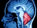
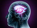
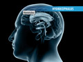
Anatomy of the Brain

The brain is the control center of the human body. It forms your thoughts and preserves your memories. It regulates your body’s actions, from the movements you choose to perform to the functions you don’t even consciously think about. Let’s take a closer look at the anatomy and the function of the brain.
Arteriovenous Malformation (AVM)
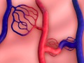
This is an abnormal connection between blood vessels. It happens when arteries connect directly to veins without first sending blood through tiny capillaries. An AVM can look like a tangle of blood vessels. They form anywhere in your body, but most often they form in or around the brain and along the spinal cord.
Astrocytoma
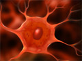
This is a tumor that begins in a brain cell called an “astrocyte.” These cells help give your brain its structure. An astrocytoma can form in your brain, in your brain stem or in your spinal cord. There are many types of astrocytomas. They can be cancerous or noncancerous. They can grow slowly or quickly. A doctor can figure out the specific type you have.
Brain Abscess

This is a pocket of pus in your brain. Tissue has grown around it, walling it off from the rest of your body. The mass is filled with white blood cells, dead tissue and germs. It can grow and press harmfully against your brain, causing a medical emergency.
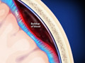
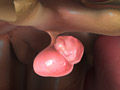
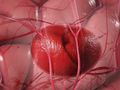
Brain Aneurysm
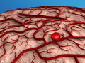
This condition is a bulge that forms in the wall of a weakened artery in the brain. This bulge can leak or rupture, causing a stroke. An aneurysm can be life-threatening.
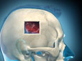
Brain Tumor (Overview)

This is a mass of abnormal cells. It may be inside your brain, or it may be next to your brain. It can grow and press harmfully against healthy brain tissue. This can cause a wide range of problems throughout your body. A brain tumor can severely impact your life.
Cerebral Cavernous Malformation (CCM)
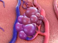
This is a mass of enlarged blood vessels in your brain or spinal cord. Pockets in the mass slow down or even trap blood. This can lead to blood clots, or to a leaking of blood we call a “hemorrhage.”
Chiari Malformation (CM)
This is a structural problem with the back of the brain. It involves the cerebellum. That’s the part of your brain that controls balance. Normally, the cerebellum sits in a space at the base of the skull. It’s just above the opening to the spinal canal, called the “foramen magnum.” With Chiari malformation, the cerebellum slips down through this opening.
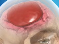
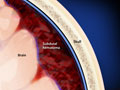
Chronic Subdural Hematoma (Hemorrhage)
This condition is a buildup of clotted blood between the brain’s outer layer and the membrane that covers the brain (called the dura). It usually occurs in the elderly, and can be caused by even a minor bump to the head.
How Your Brain Changes With Age

Like every part of your body, your brain changes as you age. And some changes affect how you think. Let’s look at what’s normal, and let’s talk about things that may be cause for concern.
Hydrocephalus

This condition is caused by an increased amount of cerebrospinal fluid (commonly called CSF) in the brain’s ventricles. The ventricles are a system of large, fluid-filled open spaces inside the brain. Too much CSF in the ventricles can elevate pressure in the skull. It can damage delicate brain tissue.
Meningioma
This is a tumor in your meninges. These thin layers of protective tissue surround your brain and spinal cord. Most meningiomas are not cancerous. They usually grow slowly.
Metastatic Brain Tumor
This is a cancer that began elsewhere in your body and then spread to your brain, forming one or more tumors. Many different cancers can spread this way. These tumors are actually more common than tumors that begin in the brain’s own tissues.
Myelopathy

This is a problem that affects your spinal cord. It happens when something presses harmfully against it. Your spinal cord is the main nerve pathway between your brain and your body. Pressure on it can cause problems throughout your body.
Normal Pressure Hydrocephalus (NPH)
This condition, which usually occurs in adults 55 and older, is an excessive accumulation of cerebrospinal fluid (CSF) in the ventricles of the brain. The ventricles are a system of large, fluid-filled open spaces inside the brain. Too much CSF in the ventricles can distort the brain’s shape. It can make the brain susceptible to injury.
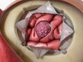
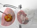
Pituitary Tumor
Your pituitary gland is found just under your brain. This pea-sized gland makes hormones that affect many of your body’s functions. A pituitary tumor can cause it to release too much or too little of these hormones. This can cause serious problems.
Pseudotumor Cerebri

This condition, sometimes called a false brain tumor, is a buildup of cerebrospinal fluid pressure in the skull. It most commonly affects obese women ages 20 to 50. The reason it develops is unknown.
Surgical Care and Management
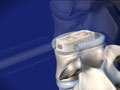
Aneurysm Clipping
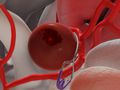
This surgical procedure is performed to treat an aneurysm, a bulge in the wall of an artery, inside the skull. Aneurysms can often become so large that they rupture or leak. In this procedure, a small, metal clip is applied to the base of the aneurysm to prevent blood leakage.
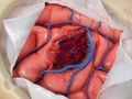
Bifrontal Craniotomy for Tumor
This surgery is used to remove a tumor from the frontal lobe of the brain. The procedure is performed under general anesthesia and requires a hospital stay.
Burr Hole Drainage
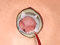
This procedure creates one or more holes in the skull to release excess fluid pressure in the brain caused by a chronic subdural hematoma (blood clot on the brain). It can be performed under local anesthesia.
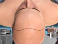
Craniectomy for Chiari Malformation (Foramen Magnum Decompression)
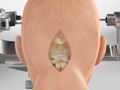
This surgery is used to treat Chiari malformation, an abnormality that results in a part of the brain extending into the upper spinal canal. During the procedure, small sections of bone are removed from the rear of the skull and spine to create more space for the errant brain tissue.
Cranioplasty
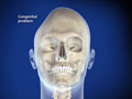
This reconstructive surgical procedure is performed to correct congenital problems of the skull, or to repair the skull after a traumatic injury or medical procedure. During the procedure, a custom plate made from porous plastic or titanium is fitted over the defect in the skull, restoring the skull to its normal shape.
Craniotomy for Epidural Hematoma
This procedure, performed under general anesthesia, creates an opening through the skull for removal of a blood clot between the skull and the dura (the membrane that surrounds the brain). Epidural hematomas commonly result from trauma to the head, and can place harmful pressure on the brain.
Craniotomy for Intracerebral Hematoma
This procedure, performed under general anesthesia, creates an opening through the skull for removal of a blood clot inside the brain. Intracerebral hematomas can result from trauma to the head. They can also occur spontaneously in patients with abnormally high blood pressure, or a blood vessel abnormality. Intracerebral hematomas can place harmful pressure on the brain.
Craniotomy for Meningioma (Brain Tumor)
This procedure, performed under general anesthesia, creates an opening through the skull for removal of a meningioma. This type of tumor is found in the dura – the fibrous membrane between the brain and skull. The surgery usually requires several hours to complete, depending on the location and size of the meningioma.
Craniotomy for Subdural Hematoma
This procedure, performed under general anesthesia, creates an opening through the skull for removal of a blood clot on the surface of the brain. Subdural hematomas commonly result from trauma to the head, and can place harmful pressure on the brain.
Craniotomy for Tumor

This procedure, performed under general anesthesia, creates an opening through the skull for brain tumor removal. The surgery usually requires between two to five hours to complete. The length of surgery depends on the type and size of the tumor.
Microvascular Decompression for Trigeminal Neuralgia
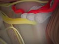
This procedure eliminates (or greatly reduces) the sharp bursts of pain in the facial nerves caused by trigeminal neuralgia. The procedure is performed under general anesthesia and requires a short hospital stay.
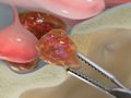
Resection of Cerebral Arteriovenous Malformation
In this procedure, performed under general anesthesia, the surgeon opens the skull to remove an abnormal tangle of enlarged blood vessels called a cerebral arteriovenous malformation (or AVM). This procedure is generally used for small AVMs that are located on or near the surface of the brain.
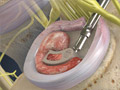
Stereotactic Biopsy
In this surgical procedure, the physician takes a small sample of tissue from the brain through a hole in the skull. Stereotactic biopsy is commonly used to take a sample from a tumor. The procedure is usually performed under local anesthesia and requires at least an overnight hospital stay.
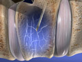
Stereotactic Radiosurgery for Arteriovenous Malformation (AVM)
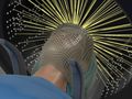
This nonsurgical procedure is used to treat an arteriovenous malformation (also called an AVM) located deep inside the brain. During this procedure, beams of radiation are precisely focused at the AVM, destroying the abnormal vessels while leaving surrounding tissue unharmed. The procedure may take several hours.
Transsphenoidal Surgery for Tumor
This endoscopic procedure, performed under general anesthesia, is used to remove a tumor from the pituitary gland. The patient will require hospitalization for two to five days after the surgery.
Peripheral
Conditions
Bursitis of the Hip (Trochanteric Bursitis)
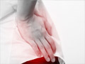
This is an irritation or swelling of the trochanteric bursa. This small, fluid-filled sac is found on the outer side of the femur. It acts as a cushion for the iliotibial band, a thick tendon in your leg.
Complex Regional Pain Syndrome (CRPS)

This is a type of chronic, long-lasting, pain. In most cases, it develops in an arm or a leg that you have previously injured. With CRPS, you may have unexplained pain that won’t go away. It may be severe, and it may spread.
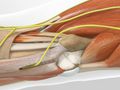
Cubital Tunnel Syndrome
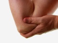
This condition, also called “ulnar nerve entrapment,” happens to the ulnar nerve in your elbow. This nerve travels along the inner side of your elbow and down to your hand. It’s the nerve that makes the jolt you feel when you bump your “funny bone.” With this condition, your ulnar nerve is compressed, stretched or irritated.
Spine
Conditions
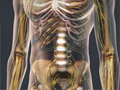
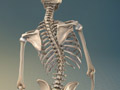
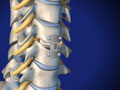
Anatomy of the Spine
The spinal column is the body’s main support structure. Its thirty-three bones, called vertebrae, are divided into five regions: cervical, thoracic, lumbar, sacral and coccygeal.
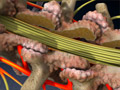
Cervical Radiculopathy
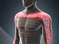
This condition is an irritation or compression of one or more nerve roots in the cervical spine. Because these nerves travel to the shoulders, arms and hands, an injury in the cervical spine can cause symptoms in these areas. Cervical radiculopathy may result from a variety of problems with the bones and tissues of the cervical spinal column.
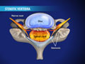
Compression Fractures of the Spine
This is a collapse of vertebral bone. It can affect one or more vertebrae. Compression fractures typically develop in your mid or lower back. This can change the shape of your spine.
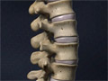
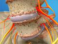
Degenerative Disc Disease
This condition is a weakening of one or more vertebral discs, which normally act as a cushion between the vertebrae. This condition can develop as a natural part of the aging process, but it may also result from injury to the back.
Facet Joint Syndrome
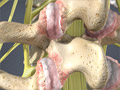
This condition is a deterioration of the facet joints, which help stabilize the spine and limit excessive motion. The facet joints are lined with cartilage and are surrounded by a lubricating capsule that enables the vertebrae to bend and twist.
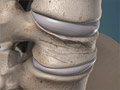
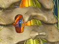
Herniated Disc (Cervical)
This condition is a rupture of one of the vertebral discs in your neck. A herniated disc can allow disc material to press harmfully against the spinal nerves.
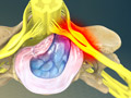
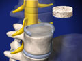
Herniated Discs
A herniated disc is a common injury that can affect any part of the spine. A herniated disc can cause severe pain and other problems in the arms or legs.
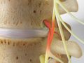
Lumbar Radiculopathy (Sciatica)
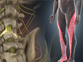
This condition is an irritation or compression of one or more nerve roots in the lumbar spine. Because these nerves travel to the hips, buttocks, legs and feet, an injury in the lumbar spine can cause symptoms in these areas. Sciatica may result from a variety of problems with the bones and tissues of the lumbar spinal column.
Osteoarthritis of the Spine

If you have back or neck pain that doesn’t go away, you may have osteoarthritis of the spine. Osteoarthritis is the most common form of arthritis. For many of us, it develops slowly as we age. And it can keep you from being as active as you like.
Post-Laminectomy Syndrome

This condition, also called “failed back syndrome,” is a type of chronic pain. It can develop in some people after spine surgery.
Scoliosis
This condition is an abnormal curvature of the spine. It most often develops in early childhood, just before a child reaches puberty.
Spinal Stenosis (Cervical)
This condition is a narrowing of the spinal canal that results from the degeneration of bones, discs, or joints in the cervical spine.
Spinal Stenosis (Thoracic)
This condition affects the thoracic spine between the neck and the lower back. It is a narrowing of the spinal canal that results from degeneration of bones in the spine, disc herniation, or thickening of the tissues that surround the spinal cord.
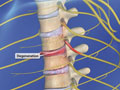
Spondylolisthesis
This condition occurs when a lumbar vertebra slips out of place. It slides forward, distorting the shape of your spine. This may compress the nerves in the spinal canal. The nerves that exit the foramen (open spaces on the sides of your vertebrae) may also be compressed. These compressed nerves can cause pain and other problems.
Where Lower Back Pain Begins
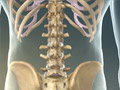
Lower back pain is a common problem that severely impacts the quality of your life. It can limit your ability to be active. It can cause you to miss work. Many different causes may lead to pain in your lower back.

Surgical Care and Management
ALIF: Anterior Lumbar Interbody Fusion (with bone graft and pedicle screws)
ALIF is generally used to treat back or leg pain caused by degenerative disc disease. The surgeon will stabilize the spine by fusing vertebrae together with bone graft material.
Anterior Cervical Corpectomy
This surgery relieves pressure on the spinal cord and the spinal nerves. It involves the removal of bone and discs from your cervical spine, followed by a fusion.
Anterior Cervical Discectomy and Fusion (Intervertebral Spacer)
This surgery removes a herniated or diseased disc and relieves neck and radiating arm pain caused by parts of the disc pressing on nerve roots.
Artificial Cervical Disc Replacement (Prestige®)
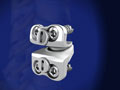
This procedure replaces a diseased or damaged spinal disc with a specialized implant designed to preserve motion in the neck. This procedure can relieve the pain of pinched nerves in the cervical spine.
Cervical Posterior Foraminotomy
This surgery creates more space for a compressed spinal nerve in your neck. The procedure relieves painful pressure caused by a herniated or degenerative disc.
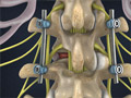
Laminectomy
This procedure relieves pressure on the nerve roots in the spine. It is most commonly performed to relieve the pain of stenosis. This is a narrowing of the spinal canal that is often caused by the formation of bony growths that can press against the nerve roots. The surgeon may treat one or more vertebrae.
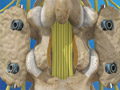
Laminectomy (Cervical)
This procedure removes a section of bone from the rear of one or more vertebrae to relieve the painful and disabling pressure of stenosis.
Laminectomy (Cervical) with Fusion
This procedure removes a section of bone from the rear of one or more vertebrae to relieve the painful and disabling pressure of stenosis. The spine is then stabilized with rods and screws.
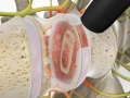
Lumbar Disc Microsurgery
This minimally-invasive procedure relieves pressure on nerve roots caused by a herniated disc. It can eliminate the pain of sciatica.
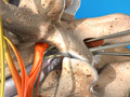
Minimally-Invasive Lumbar Microdecompression
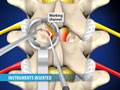
This minimally invasive procedure is used to remove overgrown vertebral bone and soft tissue to relieve the compression of nerve roots in the lumbar spine. It is performed through a small incision on the back.
Minimally-Invasive TLIF (Transforaminal Lumbar Interbody Fusion)
This minimally invasive procedure is used to remove a degenerated disc to relieve the compression of nerve roots in the lumbar spine. It is performed through a small incision on the back.
OLIF: Oblique Lumbar Interbody Fusion (for L2-L5)
This is a surgery to correct problems caused by a degenerated disc in your spine. It creates more space for your nerves. OLIF is performed through a small opening in your side.
Spinal Cord Stimulation (Paddle Lead)
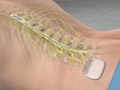
Spinal cord stimulation (also called SCS) uses electrical impulses to relieve chronic pain of the back, arms and legs. It is believed that electrical pulses prevent pain signals from being received by the brain. SCS candidates include people who suffer from neuropathic pain and for whom conservative treatments have failed.
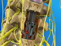
Spinal Fusion (Lumbar)
In many spinal surgeries, two or more vertebral bones are permanently joined with a technique called “spinal fusion.” A fusion creates a solid mass of bone. It stabilizes your spine.
TLIF: Transforaminal Lumbar Interbody Fusion
TLIF is generally used to treat back or leg pain caused by degenerative disc disease. The surgeon will stabilize the spine by fusing vertebrae together with bone graft material.
Vertebroplasty
This minimally-invasive procedure is an injection of bone cement into a vertebra. It stabilizes a compression fracture of the spine. One or more vertebrae may need to be treated.
XLIF® Lateral Lumbar Interbody Fusion
Unlike traditional back surgery, XLIF® is performed through the patient’s side. By entering this way, major muscles of the back are avoided. This minimally-invasive procedure is generally used to treat leg or back pain caused by degenerative disc disease. It can be performed on an outpatient basis.
Built with by Globalware.